What if every tissue sample that every pathologist in the world has ever examined under a microscope could be digitally indexed and archived? The resulting database – hundreds of millions of slides – would amount to a massive, powerful tool for medical researchers and doctors worldwide.
That very tool is now under construction. Huron Digital Pathology, a St. Jacobs’-based maker of digital imaging scanners, has partnered with the University of Waterloo and the Grand River Hospital to create the mother of all medical image search engines, fuelled by Artificial Intelligence.
The project has been underway since last May, in part thanks to a $3.1 million in funding from the Ontario Research Fund.
“It’s a huge boon to researchers. It’s very much an enabling technology,” says Patrick Myles, Huron Digital CEO.
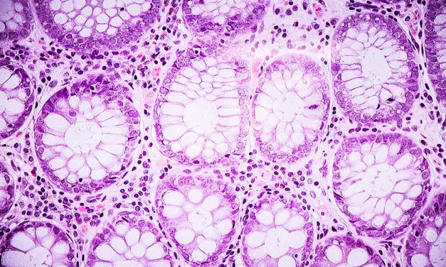
The aim of the image database project is to permit a physician or researcher to call up the magnified image of a tissue sample, and then allow the same person to search for and access a digital version of other samples that might be similar or related. The images can then be compared.

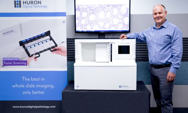
Huron Digital Pathology CEO Patrick Myles, with one of his company’s digital scanners.
(Communitech photo: Sara Jalali)
“It quickly accelerates very basic research that just wasn’t doable when you were looking under a glass slide,” says Myles. “You look at a glass slide, [then] you look at another one, you can’t remember what you saw on the first one. Whereas, if you can have both of them up on a screen digitally, or an algorithm that can compare them and do some analysis on them … it allows them to accelerate what would [otherwise] take years.”
The technology additionally allows medical personnel access to the images no matter where in the world they happen to be located.
“We have a customer in Puerto Rico who spends a lot of time in the south of France,” sayd Myles. “He can have the processing of the tissue done in Puerto Rico and he can look at the images in France on a cell phone or tablet or laptop. Wherever you have internet connection you can do the work.
“It speeds things up. You don’t have to hop in a car go to the hospital. You can look at things remotely.”
The scanner used to create the images is, in effect, “a highly automated digital microscope” which can be loaded with up to 120 slides of tissue samples. With the press of a few buttons the scanner creates a high-resolution image of the slides that can be viewed on a monitor and shared.
It’s hoped that the emerging database will, in particular, speed the diagnoses of cancers.

Magnified tissue sample produced by Huron Digital Pathology’s digital scanner.
(Communitech photo: Sara Jalali)
“Millions of medical images are stored in the archives of hospitals and clinics, and generally include detailed patient-specific information such as biopsy findings, treatment plans, and follow-up reports,” Dr. Hamid Tizhoosh, a professor with the University of Waterloo’s Faculty of Engineering, said in a recent Grand River Hospital blog post about the database’s creation. “The ability to correlate new patient images with past diagnosed cases can assist experts to avoid [overlooking] malignancies.”
Dr. Tizhoosh is head of UW’s Laboratory for Knowledge Inference In Medical Image Analysis (also known as the KIMIA Lab) and one of the key partners on the project, along with Dr. Adrian Batten, Joint Chief of Laboratory Medicine at Grand River Hospital and St. Mary’s General Hospital.
Huron Digital has a more-than 20-year pedigree, itself emerging out of University of Waterloo. Its scanners are designed and manufactured in-house, in a purpose-built, 5,700-square-foot facility recently completed in St. Jacobs. Depending on the model and configuration, the scanners are worth between US$75,000 and US$150,000.
The company, which recently held an open house, has 21 employees and plans are in place to scale.
“We’ve always known we have something special,” said Myles. “Now we’re getting that traction. We’re bringing in AI to add that smart component to what we do.”

